

From the time you get up this morning to the time you go to sleep, your jaw muscles will activate about 1500 times to chew and process food. These muscles are relevant to many of our daily activities, from eating to creating words and even exercising. But, have you ever given thought to what muscles control your jaw or how they do it? In this article, we will provide a brief overview of mastication- the process of opening and closing our jaws for eating!
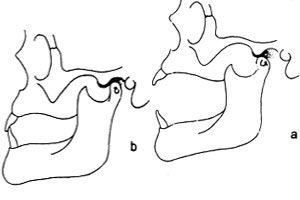
The motion of the condyle and the TMJ. Notice how it moves both forward and rotated downward when transitioning from (b) to (a).
When we open our mouths, a complex set of motions occur at the temporomandibular joint. Essentially, the condyle of the mandible (jaw bone) fits into a specialized groove in the skull. When muscles are activated to open the jaw, the condyle moves forward and out of this groove while rotating open. This movement allows a greater range of motion for chewing and swallowing. Unfortunately, this intricate machinery can become damaged or displaced. Some patients with histories of grinding or clenching their teeth will develop Temporomandibular Joint Disorder (TMJD).
Although there are a number of muscles that assist and aid in the opening and closure of the jaw, there are four main muscles whose sole purpose is mastication. The masseter, temporalis and medial and lateral pterygoid muscles are all uniquely dedicated to making eating possible. Interestingly enough, all of these muscles are controlled by the same nerve- the mandibular branch of the trigeminal nerve. Together, they allow us to open our jaw, move it left and right and close it with incredible force.
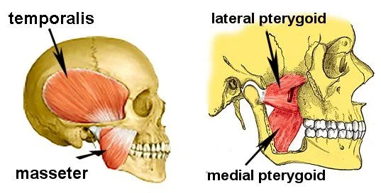
The muscles of mastication labeled. In the second image, the first masseter, temporalis and part of the jawbone have been removed.
The masseter muscle runs from the side of the skull down to the bottom of the jawbone. If you place your hand under your temple and before your ear, you can feel it tense as you bite down. It’s main purpose is to help close the jaw. If you follow the masseter around the underside of your jaw, it makes a sling with the medial pterygoid muscle. This muscle also helps close the jaw, along with moving it from side to side. The lateral pterygoid sits just outside of the medial pterygoid and opens the jaw while assisting in side to side motion as well. Finally, the temporalis muscle sits along the side of the skull and is composed of two different types of muscle fibers. The vertical fibers (running top to bottom) help close the jaw, while the horizontal fibers (running from front to back) are responsible for retracting the jaw.
Each of these motions is crucial to how we use our jaws in daily life. Particularly in chewing, these complex movements allow us to properly process our food before it reaches our digestive tracts. If you were keeping tally, we have three muscles dedicated to closing the jaw, and only one that functions in keeping it open (lateral pterygoid). This gives us some insight into how incredibly important proper chewing function is to our overall health. If you would like to know more about the muscles of mastication, facial anatomy or any dental topics, please contact our office. We want our patients to be both excited and well informed on topics in oral health!