Viewing entries in
New Technology

Dental Questions: What are digital x-rays?

Comment

Dental Questions: What are digital x-rays?

thousandoaksfamilydentistry.com

Most of our patients will remember the transition from film to digital dental x-rays with a positive stance. Gone are the days of sharp film holders, waiting for the developer and difficulties in duplication. But what are the biggest advantages of digital x-rays? Are they safe? How to they work? Read on to find out the true benefits behind this new technology!

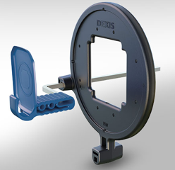
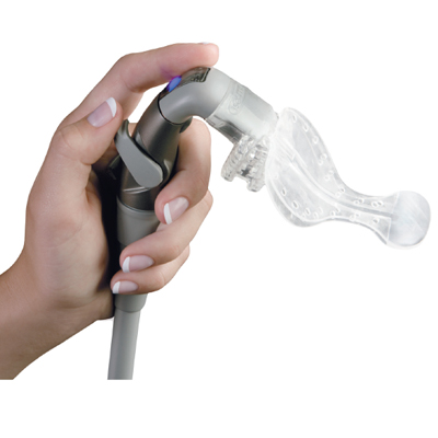
Digital dental x-rays work on the same principles as modern digital cameras. In fact, our x-ray sensors use the same CMOS technology found in many modern DSLR, pocket and cell phone cameras. The main difference is that dental sensors are calibrated to react to x-ray radiation rather than the photons in visible light. Beyond that, the images are digitally decoded and rendered using much of the same technology as digital photography. The sensor itself contains no hazardous material and emits no radiation. 

The two main principles that drive the digital revolution in dental x-rays are lower radiation and increased efficiency. "Exposing" a digital x-ray sensor requires less radiation than a similarly sized film, even when comparing the most sensitive film available. Digital images are also easier to manipulate, enlarge and change contrast, which allows us to gather more information off of a single picture. Finally, we can now judge the need for re-takes immediately, which translates to less waiting during your appointment. 

Whenever discussing dental x-rays, the topic of safety always comes to light. We are here to assure our patients that dental radiography is extremely safe, with the benefits largely outweighing any drawbacks. Even after a full-mouth set of x-rays (taken about once every five years), you experience roughly the same amount of radiation as a trans-atlantic flight. Furthermore, four bitewings (taken about once every year to 18 months) require less radiation than spending a day outside in the sun. Particularly with our ultra sensitive digital sensors, your effective radiation exposure is negligible

It is important to note that traditional film x-rays still play an important role in dentistry. Many practitioners still rely on film with excellent results and high patient satisfaction. This is particularly true of pediatric offices, where the thin profile and gentle flex of film allows dentists to get clearer images in smaller mouths. If you would like to know about digital x-rays or any other new trends in dental technology, please give our office a call!

Comment

Stimulating Stem Cells With Dental Fillings

2 Comments

Stimulating Stem Cells With Dental Fillings

thousandoaksfamilydentistry.com

You have probably seen headlines across the internet discussing new a newly invented dental filling that stimulates stem cells within the teeth. Researchers at The University of Nottingham and Harvard University came together and created this material that is proposed to change the face of dentistry. While this discovery has huge implications for dental care, many articles oversell its proposed abilities. 

In discussing how this material is supposed to work, it is important to understand the anatomic structure of teeth. Every tooth has three general layers: enamel, dentin and pulp tissue. The enamel is mostly mineral and happens to be the hardest structure in the human body. It is also the layer most resistant to tooth decay. Beneath the enamel lies the softer and porous inner dentin. The pulp tissue is the innermost layer that contains nerves, blood supply and stem cells. These new fillings rely on changing the pulp's response to trauma and inflammation. 

Unfortunately, dental pulp tissue is not very resilient. Trauma, extreme temperatures, deep fillings and bacteria can all cause the pulp to become inflamed and/or die, necessitating a root canal. However, if the attack is moving slowly enough, the pulp may shrink itself by laying down new dentin and "move away" from the irritating source. The new filling material hopes to capitalize on this mechanism when placing deep fillings near the pulp tissue. Instead of causing the nerve to die, the substance hopes to induce the tooth to lay down more dentin, creating a safe and natural barrier between the tooth and filling. 

It is important to understand that (by design) this material is not an end-all solution to cavities and root canals. For starters, it would have little benefit over traditional fillings when used in shallow cavities far from the pulp. It would also not be of use in teeth that need root canals due to fracture, trauma or decay that extends entirely to the pulp. Many times, teeth need root canals not due to deep fillings that aggravate the pulp tissues, but rather because the bacteria have travelled through the enamel and dentin and have infected the inner tooth. Once the pulp comes in contact with outside microbes, it will almost certainly need a complete root canal treatment. 

Still, this new material has wonderful applications in the modern dental office. Many fillings that were previously given with a questionable prognosis can now be provided with increased likelihood of success. Likewise, this invention paves the road for future regenerative dental technologies. Who knows what the future holds in store! For more information on these new fillings and how they work, feel free to give our office a call or email us at staff@thousandoaksfamilydentistry.com


2 Comments

Cone Beam CT Technology

Comment

Cone Beam CT Technology

Here is a series of images assembled using CBCT technology. They are being used to visualize a tooth treated with a root canal. 

Here is a series of images assembled using CBCT technology. They are being used to visualize a tooth treated with a root canal. 

Dental X-rays are very useful in diagnosing a number of conditions. Decay, abscesses and bone structure can easily be seen using standard technology found at any dental office. However, X-rays are inherently limited: they can only show you a two dimensional image of a three dimensional object. In many instances, X-rays can visualize part of a problem, but leave out "the whole picture." How deep is that crack? Which tooth is draining to that abscess? Are there one or two canals in that root? For these questions, many dentists and specialists use Cone Beam CT technology

Cone Beam CT (CBCT) is based on a principle called Computerized Tomography. Here, a series of low-radiation exposures are used to take "slices" that can assemble to a 3D image. Imagine looking at a apple, cut widthwise many times. If you saw each slice consecutively, you could easily surmise the 3D shape of the original apple. This is the same idea behind CBCT- many individual images are used to create a 3D representation of hard tissue. While a traditional X-ray might miss small problems (like a cracked root) or misrepresent sizes or shapes (like superimposing two root canals as one), a CBCT excels at giving us a more complete picture.

Many of the specialist we refer to utilize CBCT technology. For example, Doctors Bollinger, Cobin and Stoop at Conejo Simi Endontics use it to diagnose cracks, visualize root canals and determine the prognosis of treated teeth. CBCT is particularly useful for endodontics, as many problems are difficult or impossible to locate, even with high powered microscopes. Likewise, Dr. Wilgus at Camarillo Periodontics uses his CBCT machine to plan implant placement. Being able to see the height and width of bone along with any associated structures adds extra predictability to his surgeries. 

It's easy to see how CBCT technology has made diagnosing dental conditions easier and more reliable. When we make referrals to our specialist network, we have these capabilities in mind- that these doctors have the technology to visualize problems that normal X-rays can't. If you would like to know more about Cone Beam technology and how it is used in dentistry, just give us a call!


Comment

Laser Dentistry

Comment

Laser Dentistry

A dental laser in action.

A dental laser in action.

One of the biggest developments in modern dentistry is the introduction of laser technology in the treatment of patients. 50 years ago, the idea alone would have sounded like something out of a sci-fi movie. But today, lasers have many applications in the maintenance of the teeth and gums, along with promising outlooks for the future. In our office, we use lasers for a number of treatments and want to show you why they are safe and effective dental instruments!

At it’s very core, a laser is specially concentrated beam of light that produces a small pinpoint of energy. Depending on the type of material used to create the laser beam, the energy can be optimized to cut a number of materials. For this reason, lasers have long been used in precision manufacturing for making extremely accurate cuts. In dentistry, these properties allow us to use lasers in a variety of procedures, often with less time and tissue damage involved.

The most common application of lasers in dentistry is the removal or recontouring of gum tissue. In treatments where the gums need to be adjusted, a dentist would classically utilize a sharp instrument like a scalpel. While these did a great job of cutting tissue, they would induce considerable bleeding and inflammation. Lasers match the cutting abilities of standard instruments, but also stimulate healthier healing patterns from your immune system. Because the laser “seals” the tissue as it cuts, there is less trauma to the gum tissue and shorter healing times.

At our office, we particularly capitalize on the incredible healing properties of lasers in restoring dental implants. After an implant is placed and has healed in bone, it needs to be “revealed” by cutting away any gum tissue that would interfere with attaching the crown. Traditionally, we would need one appointment to trim the gums and a second appointment to allow them time to heal before taking impressions for the lab. However, our laser system allows us to reveal the implant and take impressions in the same appointment, as the gums will heal right away with minimal inflammation.

Beyond soft tissue adjustment, lasers are utilized to provide a number of dental services, ranging from stimulating periodontal attachment to removing tooth decay. Research has also shown that lasers hold even more applications for the future. Currently, experts are working on developing a system called selective ablation, where hard tooth structure is treated by a laser that only removes demineralized areas. Although this technology is still in development, it promises a new form of extremely conservative cavity removal, as only decayed tooth will be affected by the laser.

The field of laser dentistry is still new and evolving. Likewise, information and products are being introduced to the market at a rapid pace. As with all new technologies, our office likes to evaluate the research and clinical results before incorporating them into our patient treatment. If you would like to know more about dental lasers, the types of lasers we use or why we use them, please give us a call. We pride ourselves in being knowledgeable in all the current topics in dentistry and dental services!


Comment

New Technology- DEXsheild X-Ray Sheild

Comment

New Technology- DEXsheild X-Ray Sheild

The DEXsheild is a black ring that focuses the radiation produced by an X-Ray beam to the exact dimensions of our digital sensors.

The DEXsheild is a black ring that focuses the radiation produced by an X-Ray beam to the exact dimensions of our digital sensors.

When receiving medical, dental or pharmaceutical care, safety is everyone’s main concern. Adverse reactions, side effects and allergies can all make a treatment undesirable or unrealistic for your personal needs. As such, our office is always looking for new ways to minimize any potential risks while maximizing our patients’ wellness. Most recently, we have updated our X-Ray safety protocol to include the new DEXsheild radiation shield.

For starters, it is important to note that dental X-rays are an extremely safe and useful diagnostic tool. A standard dental X-ray produces roughly a quarter of the radiation you would receive on any given day. Yearly, it represents a negligible fraction of radiation exposure that our bodies absorb from the sun, consumer electronics and other sources. At our office, we further minimize any risks by utilizing high sensitivity digital X-ray sensors, radiation aprons with thyroid collars and modern X-ray beams. In short, we make sure every patient is safe and protected to standards exceeding those laid out by the ADA and other governing bodies.

The DEXsheild fits in to our current safety standards by narrowing the beam of radiation that the patient is exposed to. We have always used devices called collimated X-ray beams. Here, a thick metal tube essentially focuses the radiation produced by the beam to the oral region. While the sensor can detect and develop an image, the rest of your body is largely unexposed to any X-rays. The DEXsheild works by taking the place of a standard targeting ring (the red, blue and yellow rings we have on the outside of the sensor holders) and blocking the released radiation to the exact dimensions of the sensor. It essentially functions as a secondary safety apron, but for your mouth!

X-rays represent an indispensable tool at any dental office. The information provided from a few quick photographs can give us fantastic perspective into if a tooth is healthy, why it might be hurting or what we can do to repair it. We also understand that many patients are concerned about radiation exposure and want to know how we are keeping them safe. If you have any questions or concerns on ANY of our safety protocols, please give us a call. Our patient’s safety is our primary concern and we are happy to alleviate any of your worries!

Comment

New Technology- The Isolite

Comment

New Technology- The Isolite

Throughout the years, the practice of dentistry has certainly gone through many changes. New technologies have made the average appointment, quicker, more comfortable and more productive. At Thousand Oaks Family Dentistry, we stay at the forefront of the dental industry, always updating our equipment and techniques to the latest standards. One such instrument we are proud to utilize is the Isolite vacuum system.  

In the past, even simple dental work was clinically challenging to keep dry and clean.  An assistant would have to hold multiple suctions for the duration of your appointment, taking care to protect your tongue and cheeks. While effective, this process is cumbersome, difficult to use on certain teeth and ultimately less relaxing for the patient. The Isolite streamlines this entire process, while adding an extra dimension of comfort to your visit.

An Isolite handpiece. The clear, flexible plastic forms a mouthpiece that keeps you safe and comfortable. 

An Isolite handpiece. The clear, flexible plastic forms a mouthpiece that keeps you safe and comfortable. 

The Isolite is a specialized suction system mated to custom sized mouthpieces. It connects to the central vacuum line at our office and is installed in every one of our operatories. The mouthpieces provide a comfortable place for you to rest your mouth along with continuous suction and lighting. They have two flexible “wings” that wrap around your tongue and cheek, creating a safe barrier between our instruments and your soft tissue. The end result is a quicker, safer and more comfortable appointment. Additionally, since the Isolite provides more thorough suction, it keeps our work area cleaner and drier. This translates to more precise preparations and stronger cement bonds with better longevity. 

An example of the Isolite in use. 

An example of the Isolite in use. 

On top of coming in five different sizes, the Isolite mouthpieces can be further trimmed and shaped in a matter of seconds. Most patients find the fit to be quite comfortable, often forgetting we are using it. Overall, we have had excellent results with this system and use it for nearly every (non-cleaning) appointment. If you would like to learn more about the Isolite and why we use it, please give us a call. We are always happy to educate our patients on the instruments and techniques used at our office!

Comment

Viroxyn Cold Sore Treatment

Comment

Viroxyn Cold Sore Treatment

Viroxyn is packaged in a convenient vial/swab combination and can be applied in a matter of seconds. Credit: Quadex Labs

Viroxyn is packaged in a convenient vial/swab combination and can be applied in a matter of seconds. Credit: Quadex Labs

Every year, millions of Americans develop one or more cold sores on their lips. These blisters are unsightly, painful and contagious- often causing us to avoid or limit socialization while they are inflamed. Fortunately, there is an excellent medical solution to this pain and embarrassment. Our office is proud to offer Viroxyn- a professional strength cold sore treatment.

Cold sores are caused by a viral infection that enters the body via the mucosal tissue (i.e. lips) or dry/cut skin. Once inside, it can lie dormant for months or years, until it becomes activated and begins to replicate. As the virus grows, your body’s immune system kicks in and begins to supply the area with white blood cells to combat the infection. This process causes the typical cycle of pain, irritation and appearance of blisters associated with cold sores.

Untreated, the entire biological process takes about 2 weeks to resolve itself. The virus then becomes dormant until reactivated, as it is never actually eradicated from your body. Typical cold sore sufferers receive an average of 1-3 outbreaks a year, often coinciding with times or stress or irritation to the lip tissue. Dry weather, hectic work schedules and even sunburns can cause the virus to engage and begin replicating.

Viroxyn helps minimize the hassle of these outbreaks through a combination of an effective pain reliever and antiseptic. One applied, the topical anesthetic begins to numb the pain, while the antiseptic helps the body target and eradicate the active virus. Many patients find their pain eliminated in one hour, with the visible outbreak minimized in as few as three days. Viroxyn works best when the outbreak is caught early on, and is typically administered as soon as you feel the first signs of a cold sore. The medicine is delivered in a convenient single use swab system and takes less than a minute to apply. 

If you suffer from cold sores, please let us know at your next visit. This product does an excellent job of stopping pain from cold sores and can drastically shorten your time spent suffering with an unsightly blister. If you have any further questions about this product, or would like to learn more about what causes cold sores, please call our office. We are here to help in every way that we can!

Comment

Kerr Sonicfill Filling System

Comment

Kerr Sonicfill Filling System

If you have had a filling placed at our office recently, you likely noticed that the entire appointment was much shorter (and more comfortable) than when you were a child. This is due to a number of wonderful advancements in dental technologies and techniques, allowing us to move more smoothly and efficiently. A great example of this progress is the Kerr Sonicfill Composite Applicator.

The Sonicfill "handpiece" and composite filling material tubes. It plugs into our chairs much like the dental polisher and "drills." 

The Sonicfill "handpiece" and composite filling material tubes. It plugs into our chairs much like the dental polisher and "drills." 

The Sonicfill is an incredible device that allow us to place certain fillings in the back of the mouth in a matter of seconds. Previously, properly filling a large cavity in the molars and premolars required two different types of materials, which had to be carefully packed, shaped, adjusted and finally cured. With Sonicfill, The entire process is completed in a single step, with outstanding results.

This incredible device works by utilizing the air supply built into our dental chairs to create a vibrating/pushing motion against a tube of tooth colored filling material. The vibrations cause the material to flow gently and thoroughly into the shape of the cavity, leaving no gaps or bubbles. Once the vibration is stopped, the material remains firm but pliable, allowing us to shape it perfectly before curing it completely solid. The end result is a beautiful tooth colored filling placed in practically no-time!

We love this technology for two reasons. First, it has provided us with excellent functional results. The vibrating motion truly does make a difference and has given hundreds of our patients strong, long lasting restorations. Secondly, its speed-of-use means a shorter appointment time. This translates to less time in our office, away from work and/or family.

As with all of our products, if you have any questions or concerns please call our front desk. We only use new technologies when we are absolutely sure they will provide a superior experience for our patients. Sonicfill has satisfied this requirement by making your appointment smooth, quick and comfortable!


Comment

BruxZir Crowns

Comment

BruxZir Crowns

http://www.thousandoaksfamilydentistry.com/blog/2014/6/19/bruxzir-crowns

At our office, we want all of our dental work to be beautiful, functional and enduring. Many times, finding the intersection point of these three qualities requires us to turn to new technologies and developments in dental materials. In the case of dental crowns, we utilize BruxZir milled zirconia as a beautiful restorative solution that stands up to even the toughest conditions.

In the human mouth, the farther back a tooth is, the more relative force it will receive during a regular day. Teeth closest to the jaw joint are situated right next to the masseter, the muscle responsible for chewing, biting and closing the mouth. This muscle can generate incredible amounts of force, enough to crack or fracture your own teeth. Because of this, many types of crowns will chip, break or fail in the rear of the mouth.

A diagram highlighting the masseter muscle. 

A diagram highlighting the masseter muscle. 

In the past, porcelain fused to metal (PFM) crowns were the best tooth colored solution for posterior teeth, as their solid metal underpinnings are incredibly durable. However, the baked on porcelain can fracture and break during normal wear, requiring replacement or adjustment. Gold crowns were popular as well, so long as the patient was comfortable without a tooth colored restoration. Other materials, such as full porcelain crowns, are simply too fragile to be placed in such a functional position.

In contrast, Bruxzir crowns combine longevity, strength and natural beauty. They are milled out of a solid piece of engineered zirconia to match the color and shape of the original tooth. Since it is a single piece of material, it better matches the light characteristics of natural teeth. There is no metal backbone block out and less tooth structure has to be removed to accommodate the newly made crown.

While some materials (such as Emax) offer better translucency and color properties, nothing can stand up to BruxZir’s durability. They are essential chip, grind and crack proof. Even with heavy grinders and clenchers, the single piece zirconia will not fail or erode. No other material can offer this level of endurance in an esthetic, tooth colored package.

Even taking a hammer to a BruxZir crown will not ruin it's structural integrity. 

Even taking a hammer to a BruxZir crown will not ruin it's structural integrity. 

We typically only place BruxZir crowns on molars and more rearward teeth. While these units are esthetically superior to PFMs, they are not as cosmetically versatile as Emax crowns. In essence, their incredible strength comes with the price of poorer relative translucency. For front teeth experiencing less average force and wear, we will typically place this more esthetic material. Of course, we still place PFM and gold crowns, but most patients are eligible and more willing to be fitted with these state-of-the-art units.

If you have any further questions about BruxZir crowns, or any other crown materials at our office, please give us a call. We would love to talk you through your crown options, and which material would work best for you personally!

Comment

Emax Lithium Disilicate Crowns

Comment

Emax Lithium Disilicate Crowns

    When it comes to dental crown materials, there are many different options to suit a multitude of different scenarios. Some, like full porcelain crowns, provide excellent esthetic results. Others, such as full gold units, provide extreme durability along with desirable tooth-like physical properties. However, what options offer both beautiful cosmetic results along with enduring strength?

For many years, porcelain-fused-to-metal (or “PFM”) crowns were the cornerstone of “form meets function” in crown materials. These units are made by taking an alloy metal core and attaching a thin layer of porcelain over it, resulting in a durable tooth colored restoration. Unfortunately, this process is not without it’s downfalls. The porcelain has a tendency to crack and break between the opposing tooth and the hard metal sublayer. Additionally, the alloy substructure is extremely opaque, and prevents the overall crown from having the translucency of a natural tooth. This detracts from the overall esthetics of the final product.

While PFM crown still have a very useful place in dentistry (and are still regularly placed at our office for various reasons), modern technology has provided us with newer and more elegant solutions. Today, we can offer Emax as a means of creating a beautiful crown or bridge with long term durability.

An example of a finished Emax dental bridge (right) and crown (left)

An example of a finished Emax dental bridge (right) and crown (left)

Emax is a lithium disilicate material that combines the translucency of porcelain with incredible strength. On average, these restorations can withstand 360-400 megapascals of force before breaking. What does this mean? Essentially, you could hammer a crown made of Emax into a plywood board and pull it out without damaging the structure. This incredible resilience means that Emax can be placed in almost any location in the mouth with extreme confidence.

In terms of esthetics, Emax rivals porcelain in that it provides an incredibly toothlike translucency. One of the most obvious “tells” of a dental crown is poor transmittance of light. Crown that do not respond to ambient light like natural teeth are just as visually jarring as poor tooth shade matching. Emax offers this low opacity in conjunction with the ability to have natural artifacts “baked” into the crown’s surface. Surface lines, color gradients and staining can be added to the crown to blend it perfectly with the adjoining teeth. Overall, this step helps detract from the “chiclet gum” look that is imparted to certain crown.

Of course, Emax in only one of the excellent crown options offered at our office. While it has some great properties, it can be a poor choice of restoration depending on a number of factors. Your crown material should take into account mouth location, neighboring teeth and your personal expectations. We regularly work with a number of different materials because our patients regularly need a number of different materials. If you have any questions Emax, or any other materials we offer at our office, please let us know at your next appointment!

Comment